

Introduction
Artificial intelligence (AI) represents a transformative technological development that is expected to enhance and revolutionize both clinical practice and biomedical research [1]. It encompasses intelligent systems that learn iteratively and mimic human reasoning abilities to accomplish challenging tasks [2]. Artificial intelligence (AI) has increasingly become an integral part of healthcare, offering significant benefits such as reducing the time and costs associated with drug development and enhancing the accuracy of medical diagnostics. It has also contributed to major advances in genomic analysis and the identification of disease risk factors. As a result, AI applications now span a wide range of healthcare domains, including preventive care, public health, hospital-based medicine, and surgical practice. The evolution of machine learning and deep learning technologies has enabled healthcare systems to move beyond traditional rule-based approaches toward more individualized and patient-centred care [3].
Recent advancements in AI have further increased its capabilities, raising the possibility that certain tasks traditionally performed by physicians may be augmented or, in some cases, automated. Despite these promising developments, several challenges continue to hinder the widespread adoption of AI in healthcare. One of the primary concerns is the limited transparency of many AI algorithms, often referred to as the “black box” problem. This lack of explainability can conflict with the principles of evidence-based medicine, where clinical decisions are expected to be transparent, interpretable, and justifiable [4].
Current applications of AI in Dentistry and Maxillofacial surgery
Maxillary Sinus Pathologies
Artificial intelligence (AI) is increasingly being used to detect and evaluate maxillary sinus pathologies on CT and CBCT images [6]. Deep learning models, particularly convolutional neural networks (CNNs), can automatically identify and segment conditions such as mucosal thickening and mucous retention cysts. These systems provide accurate measurements of lesion size and sinus involvement, assisting clinicians in diagnosis and surgical planning. AI-based analysis can also maintain high performance with low-dose CBCT scans, reducing radiation exposure while preserving diagnostic accuracy. As a result, AI has the potential to improve the assessment of maxillary sinus diseases and support safer, more predictable dento-maxillofacial procedures [7,8].
Oral cancer and lymph node metastasis
Artificial intelligence (AI) is improving cancer diagnosis by enabling faster and more accurate analysis of histopathological images. AI-powered digital pathology systems can examine high-resolution whole-slide images, assist in detecting cancerous changes, and support pathologists in diagnosis. By integrating clinical, anatomical, and molecular data, AI contributes to more precise and efficient cancer detection and treatment planning [9,10].
Recent AI systems can automatically detect and segment lymph nodes, reducing the need for time-consuming manual assessment and improving diagnostic efficiency. In addition, radiomics combined with machine learning techniques can extract quantitative features from CT and CBCT images to predict occult lymph node metastasis and treatment outcomes [11]. Studies have shown that models integrating radiomic and clinical data achieve excellent predictive performance, supporting personalized treatment planning. These advancements highlight the potential of AI as a non-invasive tool for improving diagnosis, prognosis, and clinical decision-making in patients with OSCC [12,13].
Mandibular Fractures
Artificial intelligence (AI) is increasingly used for the detection of mandibular fractures on CT images. Convolutional neural networks (CNNs) can automatically analyse and classify different regions of the mandible, such as the symphysis, body, angle, and condyle, to identify fracture presence. These models achieve high diagnostic accuracy and are particularly useful for detecting subtle or occult fractures, improving the speed and reliability of trauma assessment and aiding clinical decision-making in maxillofacial injuries [14].
Orthognathic surgery and dental implantology
Recent technological progress in virtual and augmented reality has created new opportunities for their application in dentistry. These technologies are predominantly employed in oral and maxillofacial surgery, with dental implantology and orthognathic surgery emerging as the most frequently reported areas of use.
Accurate implant placement is a critical factor in dental implantology, as it directly influences both functional performance and aesthetic outcomes. Virtual reality technologies have become widely adopted in implant treatment planning by utilizing preoperative cone-beam computed tomography (CBCT) data to assess implant dimensions, positioning, angulation, and proximity to vital anatomical structures. Several software platforms have been developed to support virtual implant planning and enhance surgical precision [15].
Following three-dimensional (3D) virtual planning, the treatment plan can be transferred to the clinical setting through either static surgical guides or dynamic navigation systems. The static approach involves the digital design and computer-aided manufacturing (CAD/CAM) of surgical guides that assist clinicians during implant placement.
On the other hand, the dynamic navigation allows the real time adjustment of the direction of dental implant during surgery based on the virtual preoperative planning [16].
Plastic and Reconstructive Surgery
Artificial intelligence (AI) is increasingly transforming the field of plastic and reconstructive surgery by enhancing surgical planning, improving clinical decision-making, and optimizing postoperative outcomes. Through the analysis of large datasets containing patient demographics, imaging studies, and surgical records, AI algorithms can assist surgeons in preoperative planning by predicting surgical risks, estimating procedural complexity, and identifying the most appropriate reconstructive strategies for individual patients.
Machine learning models have demonstrated the ability to predict postoperative complications, hospital readmission rates, wound-healing outcomes, and patient satisfaction with a high degree of accuracy. These predictive capabilities support personalized treatment planning and help surgeons provide more informed counselling to patients regarding expected outcomes and potential risks.
The integration of AI with robotic-assisted surgical systems has further expanded the possibilities of reconstructive procedures. In microsurgery, where precision is critical, AI-assisted robotic platforms can enhance hand stability, reduce tremor, and improve the accuracy of vessel anastomosis and tissue manipulation. Such advancements may contribute to improved surgical efficiency and better clinical outcomes.
Computer vision and deep learning technologies have also become valuable tools in aesthetic and reconstructive surgery. AI-based image analysis can objectively evaluate facial symmetry, breast volume, scar appearance, and other aesthetic parameters, reducing subjectivity in postoperative assessment. Three-dimensional imaging combined with AI enables virtual surgical planning, simulation of expected results, and improved communication between surgeons and patients.
In reconstructive surgery, AI can analyse medical images to assess tissue viability, vascular perfusion, and flap health. Real-time monitoring systems powered by machine learning algorithms can detect early signs of complications such as flap ischemia, venous congestion, or tissue necrosis, often before these conditions are recognized through routine clinical examination. Early identification allows timely intervention and may improve flap survival rates.
AI is also being explored in craniofacial reconstruction, burn management, wound assessment, and tissue engineering. Predictive models can assist in determining optimal treatment approaches, while AI-driven image analysis supports accurate measurement of wound dimensions and healing progress. Furthermore, emerging applications involving bioengineered tissues and regenerative medicine may benefit from AI-based optimization of scaffold design, cell growth, and tissue regeneration processes.
Despite these promising developments, challenges remain regarding data quality, algorithm transparency, ethical considerations, and the need for extensive clinical validation. Continued research and multidisciplinary collaboration are essential to ensure the safe and effective integration of AI technologies into plastic and reconstructive surgical practice [17].
Conclusion
In conclusion, digital dentistry, radiomics, and virtual surgical planning are revolutionizing oral healthcare by improving diagnostic capabilities, treatment precision, and clinical outcomes. Continued technological advancements are likely to further strengthen their role in delivering personalized and evidence-based dental care.
References
- de Dumast P, Mirande D, Ruet A, Varoquaux A. Current applications of deep learning and radiomics on CT and CBCT for maxillofacial diseases. Dentomaxillofac Radiol. 2024;53(2):20230245.
- Schwendicke F, Samek W, Krois J. Disruptive innovation in dentistry: what it is and what could be next. J Dent Res. 2020;99(7):752–7.
- Mohajer-Bastami A, Moin S, Ahmad S, Ahmed AR, Pouwels S, Hajibandeh S, et al. Artificial intelligence in healthcare: applications, challenges, and future directions. A narrative review informed by international, multidisciplinary expertise. Front Digit Health. 2025; 7:1644041.
- Beam AL, Drazen JM, Kohane IS, Leong T-Y, Manrai AK, Rubin EJ. Artificial intelligence in medicine. N Engl J Med. 2023;388(13):1220–1.
- Stone P, Brooks R, Brynjolfsson E, Calo R, Etzioni O, Hager G, et al. Artificial intelligence and life in 2030: one-hundred-year study on artificial intelligence; report of the 2015–2016 study panel. Stanford (CA): Stanford University; 2016. Available from: http://ai100.stanford.edu/2016-reportXu, J.;
- Wang S, Zhou Z, Liu J, Jiang X, Chen X. Automatic CT image segmentation of maxillary sinus based on VGG network and improved V-Net. Int J Comput Assist Radiol Surg. 2020; 15:1457–65.
- Jung SK, Lim HK, Lee S, Cho Y, Song IS. Deep active learning for automatic segmentation of maxillary sinus lesions using a convolutional neural network. Diagnostics (Basel). 2021;11(4):688.
- Hung KF, Ai QYH, King AD, Bornstein MM, Wong LM, Leung YY. Automatic detection and segmentation of morphological changes of the maxillary sinus mucosa on cone-beam computed tomography images using a three-dimensional convolutional neural network. Clin Oral Investig. 2022; 26:3987–99.
- Shafi S, Parwani AV. Artificial intelligence in diagnostic pathology. Diagn Pathol. 2023; 18:109.
- Lu MY, Chen B, Williamson DFK, Chen RJ, Zhao M, Chow AK, et al. A multimodal generative AI copilot for human pathology. Nature. 2024;634(8033):466–73.
- Kubo K, Kawahara D, Murakami Y, Takeuchi Y, Katsuta T, Imano N, et al. Development of a radiomics and machine learning model for predicting occult cervical lymph node metastasis in patients with tongue cancer. Oral Surg Oral Med Oral Pathol Oral Radiol. 2022; 134:93–101.
- Ariji Y, Fukuda M, Nozawa M, Kuwada C, Goto M, Ishibashi K, et al. Automatic detection of cervical lymph nodes in patients with oral squamous cell carcinoma using a deep learning technique: a preliminary study. Oral Radiol. 2021; 37:290–6.
- Wang X, Xu Z, Tong Y, Xia L, Jie B, Ding P, et al. Detection and classification of mandibular fracture on CT scan using deep convolutional neural network. Clin Oral Investig. 2022; 26:4593–601.
- Kwon HB, Park YS, Han JS. Augmented reality in dentistry: a current perspective. Acta Odontol Scand. 2018;76(7):497–503.
- Holst S, Blatz MB, Eitner S. Precision for computer-guided implant placement: using 3D planning software and fixed intraoral reference points. J Oral Maxillofac Surg. 2007;65(3):393–9.
- Barone M, De Bernardis R, Persichetti P. Artificial intelligence in plastic surgery: analysis of applications, perspectives, and psychological impact. Aesthetic Plast Surg. 2024;49(5):1637–9.
FIGURES
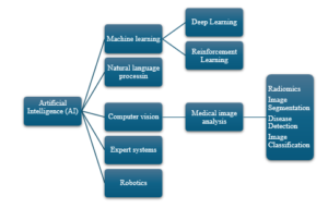
Subfields of AI